
New practices improve stroke care
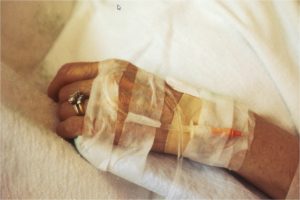
A new method of evaluating and prioritizing treatment for patients with suspected acute stroke, which has been used by the Stockholm health authority since 2017, has led to faster health interventions and better patient care, shows a new study.
Stroke can be caused by a clot in the large arteries of the brain. For every minute that an artery is blocked, two million neurons die. Without acute intervention, only 10 percent of the patients return to normal function three months after their stroke. The most effective intervention is mechanical clot removal, or endovascular thrombectomy (EVT). The earlier the blood vessel is re-opened, the greater the number of brain cells that survive. However, there is commonly some delay before treatment, as most patients are taken by ambulance to the nearest hospital for examination, and then transferred to EVT-performing university hospitals, such as the Karolinska University Hospital in Solna.
“There has been a justification for this system, as patients who don’t need EVT receive the best care at the stroke unit in their local A&E hospital,” says the study’s lead author Michael Mazya, consultant at Karolinska University Hospital and researcher at the Department of Clinical Neuroscience, Karolinska Institutet. “The challenge for ambulance staff has been to assess which patients may benefit from direct transport to a university hospital, and which can be taken to the nearest stroke unit as usual.”
To reduce the time to EVT, a new triage system has been used in the Stockholm region since October 2017. The results have now been evaluated in a new study published in the scientific journal JAMA Neurology. In patients with suspected stroke, the triage involves two steps. First the ambulance nurse tests the degree of symptom severity using the A2L2 system (where A stands for arm, and L for leg). If the patient is unable to raise his or her arm for ten seconds and the leg for five, it generally indicates a severe stroke, which often requires EVT. The second step is a telephone call to a stroke physician, to exchange additional information and decide on the optimal destination.
“The telephone consultation allows the ambulance staff and stroke physician to discuss the preliminary diagnosis,” Mazya says. “The doctor can read up on the patient’s background and activate the stroke team. This really speeds up acute management and treatment decisions once the patient arrives at the hospital.”
The results show that thanks to the A2L2 test and tele-consultation, 71 percent of patients in need of EVT are now taken directly to Karolinska University Hospital in Solna, as opposed to 28 percent before. The average time from stroke onset to EVT is now two hours and 15 minutes, which is 70 minutes faster than in the old system, 65 minutes faster than the Swedish national average, and a full 1 hour and 45 minutes faster than in international randomized studies of EVT.
Faster treatment delivery has resulted in 34 percent of EVT patients completely recovering their functional ability, compared with 24 percent in the old system, despite the fact that patients treated since the implementation of the new system have been older and had a higher average stroke severity.
“The results are very pleasing,” says Christina Sjöstrand, senior consultant in charge of stroke care at Karolinska University Hospital and researcher at the Department of Clinical Neuroscience, Karolinska Institutet. “Much of the success is thanks to a close collaboration between key stroke professionals in all of the region’s hospitals, and colleagues in pre-hospital care. We are continuing to use the new triage system throughout the Stockholm region and will be presenting more comprehensive data on patient outcomes at the World Stroke Conference in Vienna in November this year.”
Michael V. Mazya, et al., Implementation of a Prehospital Stroke Triage System Using Symptom Severity and Teleconsultation in the Stockholm Stroke Triage Study. JAMA Neurology, 2020
Women’s lifestyle changes, even in middle age, may reduce future stroke risk
Middle age may not be too late for women to substantially lower their stroke risk through lifestyle modifications. Middle-aged women who quit smoking, started exercising, maintained a healthy weight and made healthy food choices saw a reduction in their risk of stroke.
Middle age may not be too late for women to substantially reduce their stroke risk by not smoking, exercising, maintaining a healthy weight and making healthy food choices, according to new research published today in Stroke, a journal of the American Stroke Association, a division of the American Heart Association.
In general, women are more likely than men to have a stroke, die from stroke and have poorer health and physical function after a stroke. The average age of first stroke in women is 75 years. Based on this information, researchers theorized that making mid-life lifestyle changes might help reduce stroke’s burden among women.
“We found that changing to a healthy lifestyle, even in your 50s, still has the potential to prevent strokes,” said Goodarz Danaei, Sc.D., lead study author and Bernard Lown Associate Professor of Cardiovascular Health at Harvard T.H. Chan School of Public Health in Boston. “Women who made lifestyle modifications in middle age reduced their long-term risk of total stroke by nearly a quarter and ischemic stroke, the most common type of stroke, by more than one-third.”
Researchers analyzed the Nurses’ Health Study, which includes health information on nearly 60,000 women who enrolled at average age of 52 and continued in the study for an average of 26 years. Researchers studied the impact on stroke risk from smoking cessation, exercising 30 minutes or more daily and gradual weight loss if women were overweight. The researchers also studied the impact of making recommended dietary modifications that emphasize eating more fish, nuts, whole grains, fruits and vegetables and less red meat, no processed meat and less alcohol.During the 26-year follow-up, researchers found:
- 7% of women with no lifestyle interventions had a stroke of any type; 2.4% had ischemic stroke; and 0.7% had hemorrhagic stroke.
- Engaging in the three non-dietary interventions — smoking cessation, daily exercise and weight loss — was estimated to reduce the risk of total stroke by 25% and ischemic stroke by 36%.
- Sustained dietary modifications were estimated to reduce the risk of total stroke by 23%.
Researchers also found that increasing fish and nut consumption and reducing unprocessed red meat consumption appeared to have positive impacts on reducing stroke risk, although the degree of impact from these dietary changes was not as big as those achieved through increased physical activity, smoking cessation and maintaining a healthy weight.
While this was an observational study that included mostly white, middle-aged women, Danaei said, “there are other studies to support that the proportional changes in stroke risk from lifestyle and dietary modifications may be generalizable to men. We also estimate that exercising 30 minutes or more daily may reduce the risk of stroke by 20%.”
Priyanka Jain, et al.,Hypothetical Lifestyle Strategies in Middle-Aged Women and the Long-Term Risk of Stroke. Stroke, 2020
Artificial intelligence can speed up the detection of stroke
Timely detection and accurate segmentation of acute ischemic stroke (AIS) lesions on magnetic resonance images (MRIs) are essential for the triaging patient for endovascular therapy. Lesion segmentation is a routine process where the abnormal areas within brain images are qualitatively and manually picked by expert radiologists. However, manual lesion segmentation is time consuming and suffers from operator-bias. Accordingly, efficient and low-cost approaches for AIS lesion screening are yet to be introduced.
This research introduces a novel and fully automated technique for detection and segmentation of AIS lesions on MRIs and classification of images into stroke and none-stroke. This fully automated anomaly-detection method compares diffusion weighted images (DWIs) and apparent diffusion coefficients (ADC) images of the subjects with a group of healthy images in voxel-level. Areas with hyperintensity on DWI and hypointensity on ADC are identified as lesions and saved as lesion masks. The lesion segmentation method was investigated on approximately 100 cases. Since there is a risk of false lesion identification due to the artifacts, noises, and image low resolution, the lesion masks created by the method are screened and filtered via a binary classifier which either confirms that the created lesion mask contains a real AIS lesion or not. The classification performance was evaluated on about 200 MRIs.
The published results in the Journal of Neuroscience Methods show good agreement with the manually drawn lesions by experts (gold standard). The whole approach, including lesion segmentation and image classification, is straightforward, fast and does not require high computation power and memory.
“We believe that this method has the capacity to be implemented on an ordinary desktop workstation integrated into the routine clinical diagnostic pipelines of the hospitals. This approach can help the radiologists to speed up the workflow of lesion detection and to reduce the operator bias in lesion segmentation owing to the reproducibility of the method,” tells project researcher Sanaz Nazari-Farsani from Turku PET Centre.
Sanaz Nazari-Farsani, et al.,Automated segmentation of acute stroke lesions using a data-driven anomaly detection on diffusion weighted MRI. Journal of Neuroscience Methods, 2020
Analyzing patients shortly after stroke can help link brain regions to speech functions
New research from Rice University and Baylor College of Medicine shows analyzing the brains of stroke victims just days after the stroke allows researchers to link various speech functions to different parts of the brain, an important breakthrough that may lead to better treatment and recovery.
The study, “Dissociation between frontal and temporal-parietal contributions to connected speech in acute stroke” will appear in an upcoming edition of the journal Brain.
Study co-author Randi Martin, the Elma W. Schneider Professor of Psychology at Rice, worked with a team of researchers led by Tatiana Schnur, associate professor of neurosurgery and neuroscience at the Baylor College of Medicine, to evaluate the spontaneous language production of 65 stroke patients by using storytelling. For the experiment, the patients were read the story of Cinderella and were then asked to retell it.
The researchers used a well-established process to score the patients — the Quantitative Production Analysis method — and relied on 13 different measures for evaluation, including words per minute, types of words, and sentence length and formation. They found that by evaluating patients between one and 13 days post-stroke, they were able to identify how different and critical components related to language production linked up with different regions of the brain. The researchers used cutting-edge techniques to relate the brain areas damaged in each individual to the degree of their impairment on these language-production measures. Specifically, they found that retrieving words and putting them into increasingly complex sentences relied on the left temporal and parietal lobes, while producing grammatical aspects of sentences relied on the left frontal lobe.
Martin noted there are only a few other studies that have looked at stroke patients in the acute stage, but those focused on ability to produce single words rather than providing a detailed analysis of language production. The majority of studies, she said, look at stroke patients in the chronic stage of recovery, which is at least six months after the stroke. At that time, considerable reorganization of language function in the brain may have occurred. Also, studying individuals at the acute stage allows for studying those with smaller areas of damage, she said. Those with small lesions are likely to recover and thus not included in studies of chronic stroke, and examining these people allows for a more precise mapping between areas damaged and language abilities.
“Many patients in the chronic stage of stroke have significantly worse brain damage than acute patients and have plateaued with their recovery,” she said. “Their brains cannot be evaluated in the same way as acute stroke patients.”
Future work will look at these same individuals at different stages during their first year of the recovery process. One important issue will be to determine what areas of brain damage and what language abilities will predict performance a year after stroke.
Martin hopes this work will help better understand how different brain regions recover from stroke. She expects the work will be useful in the design of treatment options for stroke patients, including early interventions that may boost long-term recovery.
Tatiana T Schnur, et al.,Dissociation between frontal and temporal-parietal contributions to connected speech in acute stroke. Brain, 2020
Stroke: When the system fails for the second time
After a stroke, there is an increased risk of suffering a second one. If areas in the left hemisphere were affected during the first attack, language is often impaired. In order to maintain this capability, the brain usually briefly drives up the counterparts on the right side. But what happens after a second attack? Medical researchers have now found an answer by using virtual lesions.
It is now widely known that the brain is much more malleable than once thought. Even after stroke or brain injury the brain often succeeds finding a new balance between the failed regions and the functions they serve. Commonly, neighbouring regions are activated as well as homologues on the other side of the brain side. During language processing, the homologues of the left-dominant language areas are usually less active and are kept in check by the dominant half — until the emergency case occurs.
Until now, it was unclear whether these mechanisms also apply in the event of a second attack. Does the brain retain its capacity to adapt? This is important as up to 15 percent of those affected will have a second stroke. In addition, there was disagreement about whether an activated right brain is generally good for healing. Some studies suggest that involvement of the right hemisphere helps recovery, at least in the short term. Others had shown, however, that a loss of language areas in the left half can literally inhibit the right half. In that case the contribution of the right hemisphere has nothing to do with language and can cause confusion. The brain gets out of step. Further, studies had also found that the patients are better off if the overactive half is restrained by inhibitory magnetic stimulation. The activity is more and more shifting back to the left hemisphere. It wins the upper hand again.
Scientists at the Max Planck Institute for Human Cognitive and Brain Sciences (MPI CBS) in Leipzig, Germany, have now found that the brain areas on the right side also become more active when there is a second injury in the left language areas. “In the recovered brain, the right side’s contribution was still little after the first impairment. After the second event, in which large parts of the left hemisphere are not working anymore, its role becomes much more important,” explains Gesa Hartwigsen, research group leader at the MPI CBS and first author of the article, which has been published in the high-ranking, open access journal elife. “The second lesion increased the contribution of the right brain,” Hartwigsen said.
The scientists examined these relationships using 12 patients in whom the regions for processing properties of sound in the left hemisphere were injured. The incident had happened to them at least six months prior. Their brain had the opportunity to regenerate and adapt to the new situation. The researchers simulated the second disruption using so-called transcranial magnetic stimulation, which can be used to briefly halt certain areas of the brain through electrical stimuli. It can be used to simulate how the brain would react if certain areas actually fail due to a stroke or other events — and how this affects the ability to recognize sounds, for example. To do this, Hartwigsen and her team used a simple decision task. The participants heard the word “cat” and had to decide whether it consisted of one or two syllables. The individual impairment predicted the activation on the right side.The researchers also found that the stronger the fibre connection between the sister areas on the right side, the less the patient was affected by the interruption on the left.
“These results show that after large-scale disturbances, in which large parts of the left hemisphere no longer function as they should, the right hemisphere probably plays a beneficial role. Often, there is a lot of tissue in the left half of the brain that only works to a limited extent and needs support from the right side. “Other studies show that recovery is helped when the activated right side later down regulates itself and thus contributes to normalization on the left side,” said Hartwigsen. On the other hand, if the right half remains permanently up-regulated, healing is delayed.
Findings on how the damaged brain adapts to repeated injury could help to improve the therapy of stroke patients in the long term. “This may make it possible, at some point, to assess whether it would be more helpful to regulate specific areas up or down,” says Hartwigsen, confidently.
Gesa Hartwigsen, et al., Short-term modulation of the lesioned language network. eLife, 2020
“Timely detection and accurate segmentation of stroke lesions using MRI are essential for the triaging patient for endovascular therapy. Analyzing the brains of stroke victims just days after the stroke allows researchers to link various speech functions to different parts of the brain. Use of virtual lesions has shown effective in predicting the second stroke following the initial one. These new methods in evaluating and prioritizing treatment for patients with suspected acute stroke, are leading to faster health interventions and better patient care.”

Recent Comments